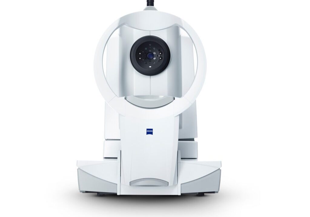
What is a Fundus Camera?

At Insight Eye Equipment, we know high-quality retinal imaging is essential for modern eye care. A fundus camera provides clear photographs of the fundus, the back surface of the eye, including the retina, macula, and optic disc. These images are a proven tool for detecting and monitoring conditions such as diabetic retinopathy, macular degeneration, and glaucoma. On this page, you’ll learn what a fundus camera is, how it supports accurate diagnosis, and why it belongs in every clinic’s diagnostic workflow.
Quick Answer: Fundus Camera Explained
A fundus camera is a specialized ophthalmic imaging device that captures high-resolution photographs of the fundus, the interior surface of the eye including the retina, macula, optic nerve head, and retinal vasculature. These images are essential for documenting eye health and play a critical role in detecting and monitoring conditions such as diabetic retinopathy, macular degeneration, and optic nerve changes associated with glaucoma.
Core Components of Modern Fundus Cameras
A fundus camera is a diagnostic imaging system designed to capture detailed photographs of the fundus, the posterior segment of the eye, including the retina, macula, optic disc, and retinal vasculature.
Key components include:
- Precision optics and mirror assembly to focus accurately on the retina
- Controlled illumination source (typically low-power flash or LED) for consistent, patient-friendly lighting
- High-resolution digital sensor that produces clear, reproducible retinal images suitable for documentation and diagnosis
How It Works: Optical Principles & Imaging Process
Modern fundus cameras apply the principles of indirect ophthalmoscopy with precision optics and digital imaging sensors to capture clear retinal photographs.
- Illumination: A ring-shaped flash or LED provides uniform, glare-free lighting through the pupil, whether dilated or undilated (in non-mydriatic systems).
- Optics: A carefully aligned lens and mirror assembly directs reflected light from the retina to the imaging sensor.
- Image capture: Fundus cameras use high-resolution digital sensors to deliver clear, color-accurate retinal images for documentation and diagnosis. Non-mydriatic models are designed to capture quality images through small pupils, helping streamline clinical workflow.
Types of Fundus Cameras
At Insight Eye Equipment, we carry a wide range of refurbished fundus cameras. Below are some of the most notable models with their key features:
- Canon CR-2 Non Myd Fundus Camera – Compact, lightweight system with LED flash and reliable non-mydriatic retinal imaging.
- CenterVue DRS – Digital Retinography System – Fully automated, touch-screen workflow; captures multiple fields with minimal operator training.
- NIDEK AFC-330 Non Myd Fundus Camera – Automated alignment and capture; designed for busy practices that need speed and consistency.
- Topcon 50DX Mydriatic Fundus Camera – Traditional mydriatic system requiring dilation; trusted for high-resolution retinal imaging.
- ZEISS VISUCAM PRO NM – Non-mydriatic color fundus imaging through pupils as small as 3.3 mm; telecentric optics for accuracy.
Clinical Applications & Benefits
Fundus cameras play an essential role in eye care by providing reproducible images that support diagnosis, documentation, and long-term management. Common applications include:
- Routine screening: Detect early signs of diabetic retinopathy, hypertensive retinopathy, age-related macular degeneration, and document optic nerve appearance in glaucoma management.
- Diagnostic evaluation: Assist in assessing retinal tears, uveitis, ocular tumors, and optic disc swelling such as papilledema.
- Disease monitoring: Compare serial images to track disease progression, evaluate treatment effectiveness, and guide clinical decisions.
- Education: Provide patients with a clear visual record of their condition and support clinical teaching.
Patient Experience: Preparation to Aftercare
The type of fundus camera used directly affects the patient’s experience:
- Preparation: Traditional mydriatic systems require dilation drops, which extend exam time and may cause temporary light sensitivity. Non-mydriatic designs reduce preparation and improve patient flow.
- During Imaging: Patients are positioned with a chin rest and guided to fixate on a target light. The system captures multiple images per eye in seconds, minimizing discomfort
- Aftercare: With mydriatic imaging, patients may experience light sensitivity until dilation wears off, often requiring sunglasses and short-term activity adjustment. Non-mydriatic units avoid this step, streamlining the visit.
Risks, Limitations & Common Artifacts
Fundus photography is a safe and reliable imaging method, but every system has technical considerations that affect image quality:
- Patient comfort: The bright flash may cause momentary discomfort, and pharmacologic dilation can add exam time and temporary light sensitivity.
- Artifacts: Common imaging artifacts include corneal reflections, motion blur from eye movement, and reduced clarity in the presence of media opacities such as cataracts or corneal haze.
- Limitations: Standard fundus cameras offer a restricted field of view compared to ultra-widefield systems. Image quality may also decline with very small pupils, though many non-mydriatic models are optimized to capture through pupils as small as 3–3.5 mm.
Final Thoughts on Understanding the Fundus Camera
Fundus imaging provides eye care professionals with a powerful tool for early detection and long-term monitoring of retinal disease. Beyond its role in patient care, the right fundus camera also improves workflow efficiency, enhances documentation, and supports clearer communication with patients.
At Insight Eye Equipment, we supply refurbished fundus cameras from trusted brands like ZEISS, Canon, NIDEK, and Topcon, each backed by warranty and white-glove delivery. Equipping your practice with the right imaging system ensures you can deliver accurate diagnostics while streamlining your daily operations. Call us today!
