Posts by Alex
What is a Microkeratome?
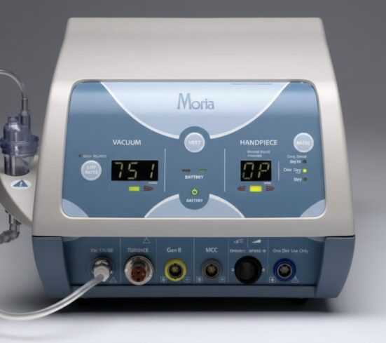
Microkeratomes still play a meaningful role in refractive and corneal surgery workflows, especially for practices that want a proven, blade based approach to flap creation and a workflow that stays consistent case after case. In this guide, we break down what a microkeratome is, how it works, where it fits clinically (including LASIK and donor…
Read MoreWhat is an Ultrawide Field Camera?

As retinal imaging advances, ultrawide field (UWF) cameras have opened new horizons for eye care.We’ve moved beyond the 10°–30° views of early fundus cameras to systems that reach the far periphery, capturing about 200° in a single image (~82% of the retina). This expanded perspective improves detection and risk stratification of peripheral disease (e.g., identifying…
Read MoreWhat is a Perimeter?

A perimeter is the instrument used for visual field testing; perimetry is the test itself that maps how you see across central and peripheral vision. Your vision extends beyond what you see straight ahead. Measuring the peripheral field is essential for detecting and monitoring glaucoma and for localizing neurologic pathway lesions (e.g., stroke, pituitary disease). …
Read MoreWhat is a Pachymeter?

Precision matters in eye care, especially when corneal thickness can influence pressure readings and surgical decisions. Pachymetry measures corneal thickness in micrometers (µm). It helps interpret applanation IOP readings, informs glaucoma risk, supports refractive surgery planning, and assists in evaluating corneal disease over time. If you’ve ever wondered what exactly a pachymeter is, it’s the…
Read MoreWhat is an OCT?
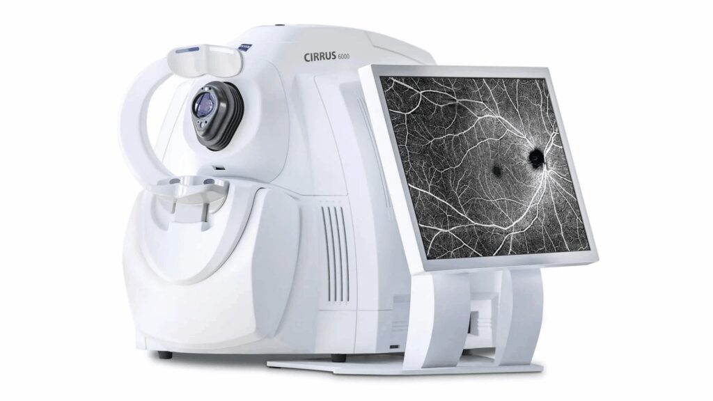
What is optical coherence tomography (OCT)? For a clear, layer-by-layer view of retinal structure, clinicians use imaging beyond standard fundus photos. Optical coherence tomography (OCT) is a noninvasive imaging technique based on low-coherence interferometry that produces high-resolution cross-sections of the eye. By mapping retinal layers and the optic nerve/nerve fiber layer, OCT helps clinicians diagnose…
Read MoreWhat is a Fundus Camera?
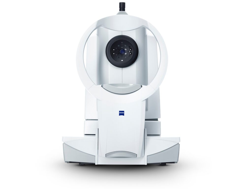
At Insight Eye Equipment, we know high-quality retinal imaging is essential for modern eye care. A fundus camera provides clear photographs of the fundus, the back surface of the eye, including the retina, macula, and optic disc. These images are a proven tool for detecting and monitoring conditions such as diabetic retinopathy, macular degeneration, and…
Read MoreWhat is a Corneal Topographer?

At Insight Eye Equipment, we know how important a healthy cornea is to clear, comfortable vision. That’s why many patients and practices turn to advanced imaging tools like corneal topographers. But what is a corneal topographer, and why does it matter for eye care? In this guide, we’ll explain the essentials and show you how…
Read MoreWhat is a Biometer?

At Insight Eye Equipment, we know precise measurements drive quality eye care. When surgeons prepare for cataract or refractive surgery, precision isn’t just a nice-to-have; it’s the difference between clear sight and ongoing uncertainty. What is a Biometer? It’s the device that provides a detailed map of your eye’s dimensions and optical qualities, guiding critical…
Read More