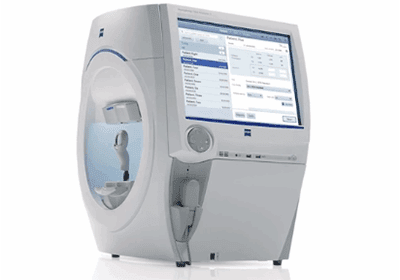
What is a Perimeter?

A perimeter is the instrument used for visual field testing; perimetry is the test itself that maps how you see across central and peripheral vision. Your vision extends beyond what you see straight ahead. Measuring the peripheral field is essential for detecting and monitoring glaucoma and for localizing neurologic pathway lesions (e.g., stroke, pituitary disease).
In this guide, we’ll walk through what perimetry is, why it matters, and how it works. You’ll learn what to expect during a test, how to interpret your results, and where visual field assessment is headed next.
Quick Answer: What Does a Perimeter Do?
A perimeter is the instrument used for visual field testing (perimetry), a test that maps how you see across central and peripheral vision. During testing, the device presents light stimuli at fixed locations while you fixate on a central target; depending on the system, this may be static (brightness changes at fixed points) or kinetic (a moving stimulus). Your button presses create a map of sensitivity, revealing defects (scotomas) that help detect and monitor glaucoma and localize neurologic pathway lesions.
Definition & Purpose of Perimetry
Understanding the visual field, central and peripheral, is essential to daily function and to clinical decision-making. Perimetry (the visual field test) measures where you can detect light while fixating on a central target, producing a map of sensitivity across your field of vision.
A perimeter (the instrument) presents light stimuli at fixed locations (static automated perimetry, e.g., ZEISS HFA) or moves a stimulus to map boundaries (kinetic perimetry, e.g., Octopus 900), charting both central and peripheral vision.
- Why full-field testing matters:
- Central vision supports recognition tasks like reading and face identification.
- Peripheral vision is critical for motion detection and spatial awareness.
- Perimetry evaluates both, helping clinicians localize defects (scotomas) and track change over time.
- Key clinical uses:
- Glaucoma — diagnosis and progression monitoring with standard automated perimetry.
- Neurologic disease — localize pathway lesions (e.g., stroke, pituitary disorders).
- Retinal disease — document central or peripheral field loss alongside imaging.
How Perimetry Works (Static vs. Kinetic)
To perform perimetry, clinics use two main testing modes, static and kinetic.
Static perimetry
- The device presents light at fixed retinal locations and varies brightness to determine threshold sensitivity (e.g., ZEISS Humphrey Field Analyzer).
- On ZEISS HFA, SITA strategies adjust stimulus intensity efficiently at each point to estimate threshold while maintaining precision.
Kinetic perimetry
- A moving stimulus maps field boundaries (classic Goldmann method). The Octopus 900 delivers full-field static and Goldmann-equivalent kinetic exams in one perimeter.
What to expect during your test
- You rest your chin/forehead on the support
- Keep fixation on a central target in the bowl
- Press the response button whenever you see a light.
- With modern SITA strategies, many exams run about 3–6 minutes per eye (protocol-dependent). For reference, published averages are ~6.2 min (SITA Standard), ~4.1 min (SITA Fast), and ~2.9 min (SITA Faster). Older/legacy strategies can take longer.
Types of Perimeters & Comparison
Clinics choose different perimeter styles based on workflow and testing goals. Here’s how the main options compare:
- Manual (Goldmann)
- Technician-guided kinetic mapping in a bowl perimeter using standardized Goldmann stimuli (sizes I–V; intensities 1a–4e).
- Technician-guided with flexible vectors and isopters, patterns can be adjusted in real time based on patient responses.
- Automated (Humphrey Field Analyzer, Octopus)
- ZEISS HFA performs static automated perimetry using SITA strategies. The Octopus 900 provides both static and Goldmann-equivalent kinetic testing in one perimeter.
- Clinic-based instruments with standardized protocols and built-in reliability/fixation monitoring (e.g., blind-spot checks, gaze tracking on HFA).
Recommended Perimeters We Carry
(All refurbished; Add to Quote to check current availability).
- Octopus 900 perimeter — Our go-to when you need both static and kinetic perimetry in one platform. The Octopus 900 maps comprehensive fields for glaucoma and neuro-ophthalmic cases without changing systems.
- ZEISS HFA3 860 perimeter — The flagship Humphrey model with the latest threshold strategies and reliability indices. The HFA3 860 streamlines standardized visual field testing for busy clinics.
- ZEISS HFA3 840 perimeter — A streamlined HFA3 configuration that delivers dependable static perimetry and modern workflow features while keeping the footprint familiar.
- ZEISS HFA 750i perimeter — A proven Humphrey analyzer that adds fixation and gaze tracking for consistent, repeatable results, ideal for longitudinal glaucoma management.
- ZEISS HFA 750 perimeter — A workhorse for routine static perimetry; the HFA 750 maintains the Humphrey testing protocols practices trust.
- ZEISS HFA 740i perimeter — Compact and clinic-friendly, the HFA 740i integrates monitoring features to keep tests reliable without slowing the schedule.
- ZEISS HFA 740 perimeter — An entry Humphrey option that provides standardized visual field testing and an accessible path into the Humphrey ecosystem.
Interpreting Results
When clinicians review a visual field test, they start with the overall maps and then confirm findings with objective indices.
- Visual field maps
- Grayscale map: A qualitative picture of sensitivity loss; useful for a quick look but not diagnostic on its own.
- Deviation plots: Total Deviation (TD) shows how each point differs from age-matched normals; Pattern Deviation (PD) adjusts TD to highlight localized loss by accounting for generalized depression (e.g., cataract).
- Key metrics
- VFI (0–100%): Global field status weighted toward central points and designed to be less affected by cataract; useful for staging and trend analysis.
- MD (Mean Deviation): Average elevation/depression of the entire field vs. age-norms; commonly used for staging and rate-of-change over time.
- PSD (Pattern Standard Deviation): Quantifies localized field irregularity; high values suggest focal loss. Less useful in very advanced loss, so lean on MD/VFI for progression there.
- Common defect patterns
- Arcuate scotoma — Nerve-fiber-bundle defect typical in glaucoma.
- Nasal step — Defect respecting the horizontal meridian (glaucoma hallmark).
- Hemianopia (homonymous or bitemporal) — Defect respecting the vertical meridian; helps localize neurologic pathway lesions (e.g., post-chiasmal vs. chiasmal).
Preparing for Your Test
If your eye care provider has scheduled a visual field test, here’s how to get ready:
- Bring your current glasses; if you wear contact lenses, bring them or your most recent prescription. The team may use a trial lens for best correction during testing.
- No special fasting or strict restrictions are required. Stay alert for the test and follow your provider’s advice about caffeine or drowsy medications the day of your appointment.
- Get a good night’s sleep so you can maintain steady fixation and respond consistently; normal hydration is fine.
- Arrive 10–15 minutes early to check in, review your prescription, and get comfortable at the instrument.
- Tell your provider about all medications (especially anything that causes drowsiness) and any recent health changes.
Final Thoughts in Understanding Perimetry
We hope this guide makes perimetry clearer and highlights why it matters for both eye and neurologic care. Whether your provider uses static or kinetic testing, knowing what to expect and how to prepare helps deliver reliable, repeatable results. If you’re curious about the latest platforms and features, explore our resources and compare models to find the right fit for your clinic. Compare perimeters, add models to your quote, or start a financing application to equip your practice with confidence. Call us today!
