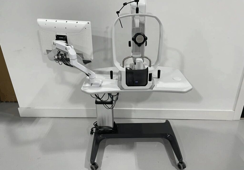
What is an Ultrawide Field Camera?

As retinal imaging advances, ultrawide field (UWF) cameras have opened new horizons for eye care.We’ve moved beyond the 10°–30° views of early fundus cameras to systems that reach the far periphery, capturing about 200° in a single image (~82% of the retina).
This expanded perspective improves detection and risk stratification of peripheral disease (e.g., identifying predominantly peripheral lesions and non-perfusion), supporting more precise monitoring and treatment planning.
Quick Answer: Understanding Ultrawide Field (UWF) Retinal Imaging
UWF cameras can capture ~200° in a single image (~82% of the retina); ~220° requires an auto-montage. By comparison, ETDRS 7-field imaging covers ~35%.
What Is an Ultrawide Field Camera?
Following the International Widefield Imaging Study Group, widefield images show retina posterior to and including the vortex vein ampullae (often ~60°–100°), while ultrawide field (UWF) images show retina anterior to the ampullae in all four quadrants (often ~110°–220°). Standard fundus photos are typically ~45° (some systems cite 30°–50° depending on lens and protocol)
- Mid-periphery: from the equator (approximated by the vortex veins) to the vortex vein ampullae
- Far-periphery: from the vortex vein ampullae to the ora serrata
Some platforms extend beyond 200° via auto-montage (e.g., OptosAdvance); true ora-to-ora 360° ‘panretinal’ composites are research-level and not typical clinical outputs.
How It Works: Core Technologies & Devices
To deliver these ultrawide views, we rely on advanced optics and sensors. You’ll see three core approaches:
cSLO systems (e.g., Optos): use multiple lasers and an ellipsoidal-mirror optical path to capture up to ~200° non-contact in a single image.
LED-based partially confocal imaging (e.g., iCare EIDON family): provides true-color images with ultra-widefield optics from ~120° up to ~200°, depending on model/configuration; some systems also support montage.
Ultra-widefield fundus cameras: ZEISS CLARUS captures 133° in a single image and ~200° with a two-image montage; RetCam (contact pediatric system) offers lenses up to ~120–130°.
Key Features & Imaging Modalities
- Color fundus photography: true-color (e.g., ZEISS CLARUS) or pseudo-color (e.g., Optos cSLO) overview of vascular and structural changes.
- Fluorescein angiography (FA) & indocyanine green angiography (ICGA): FA visualizes retinal perfusion and leakage; ICGA highlights choroidal circulation (availability depends on the device/module, e.g., Optos California with ICG).
- Fundus autofluorescence (FAF): topographic mapping of lipofuscin and related signals in the RPE, used to assess metabolic stress and disease patterns.
- UWF camera modalities: color (true- or pseudo-color), FAF, FA, and ICGA on select systems.
Clinical Benefits & Applications
Thanks to ultrawide coverage, clinicians can detect and document peripheral pathology earlier and more reliably across multiple diseases:
Diabetic retinopathy: UWF imaging identifies predominantly peripheral lesions (PPLs) in ~40% of eyes, and their presence is associated with greater severity and increased risk of DR worsening.
Retinal vein occlusion & uveitis: UWF/ UWF-FA maps peripheral nonperfusion and peripheral leakage/inflammation, improving assessment beyond standard fields.
Inherited dystrophies, tumors, sickle-cell retinopathy, ROP: UWF (color/FAF/FA) captures far-peripheral changes that may be outside ETDRS 7-field coverage, aiding diagnosis and monitoring.
Deep-learning methods on UWF images show promising performance for detecting proliferative DR features (e.g., neovascular leakage/non-perfusion), but reported metrics vary by dataset and task. In telemedicine, UWF imaging cuts ungradable rates and speeds screening workflows, so you get actionable results faster.
Final Thoughts on The Basics of Ultrawide Field Retinal Cameras
Ultrawide field cameras expand retinal visualization and documentation with 200° single-capture views, and emerging AI tools on UWF images show promise. We encourage you to assess UWF adoption based on your patient population and workflow, and to explore training resources and device-specific modules. In most clinics, a single, non-contact ~200° image (~82% of the retina) delivers the biggest lift, with ~220° via auto-montage reserved for cases where far-peripheral detail is essential.
