
Optical Coherence Tomography (OCT): What It Is and How It Works
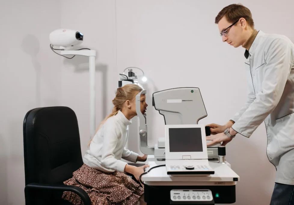
Retina specialists and comprehensive ophthalmologists rely on cross-sectional imaging to catch disease before a patient ever reports symptoms. For practices comparing a new system against a refurbished one, understanding what OCT actually measures matters as much as price. Here’s how the technology works and why it’s become standard equipment in modern eye care.
Quick Answer: What Is Optical Coherence Tomography (OCT)?
Optical coherence tomography (OCT) is a non-invasive imaging technique that uses light waves to capture cross-sectional images of the retina, optic nerve, and anterior segment. It works on a principle similar to ultrasound, but substitutes light for sound, giving clinicians micron-level resolution of tissue layers. Scans take seconds, require no contact with the eye, and produce the data clinicians use to detect and track glaucoma, macular degeneration, and diabetic retinopathy.
How Optical Coherence Tomography Captures an Image
OCT relies on a principle called low-coherence interferometry. Comparing how today’s systems execute it matters more than a single resolution number on a spec sheet, and our OCT rankings break down scan speed and software differences across the five most-installed refurbished platforms.
A light source, typically a superluminescent diode, sends a beam into the eye. Part of that beam reflects off tissue layers at different depths, and the machine compares the timing of the reflected light against a reference beam to build a depth map.
From Signal to Image
The interferometer’s output becomes what’s called an A-scan, a single vertical line of reflectivity data. Stack thousands of A-scans side by side and you get a B-scan, the cross-sectional image clinicians actually read. Modern platforms capture B-scans fast enough to build 3D volume renderings of the macula or optic nerve head in a single pass.
Spectral-Domain vs. Swept-Source OCT
Most OCT units in practices today use spectral-domain technology, which reads the interference pattern across a range of wavelengths at once using a broadband light source and a spectrometer. It’s fast, well understood, and covers the vast majority of retina and glaucoma screening needs.
Swept-source OCT, found in newer and higher-end platforms, sweeps through wavelengths sequentially using a tunable laser. That approach penetrates deeper into tissue with less signal loss, which is why it’s become the preferred choice for imaging the choroid and vitreous, structures spectral-domain systems sometimes struggle to resolve clearly. The tradeoff is cost: new swept-source platforms carry a higher price, though the gap narrows considerably on the refurbished market.
Why OCT Matters in Modern Eye Care
Before OCT became routine, clinicians leaned heavily on fundus photography and clinical exam to catch retinal disease, often after damage had already progressed. OCT changed that by making subclinical changes visible: fluid pockets from wet AMD, nerve fiber layer thinning from glaucoma, microaneurysms from diabetic retinopathy.
Most comprehensive practices now scan at every annual exam for patients over 50, and more frequently for anyone in active treatment. New to the terminology? Our OCT primer walks through the core vocabulary before you get into device specs.
OCT also extended into a related capability called OCT angiography (OCT-A), which uses the same platform to visualize blood flow in the retina without injecting dye. Where traditional fluorescein angiography requires an IV and takes several minutes to capture, OCT-A generates a comparable flow map in under a minute, noninvasively. Retina specialists use it to track diabetic retinopathy progression and detect early neovascularization, and many practices weigh OCT-A capability heavily when choosing between refurbished platforms, since not every OCT unit on the market supports it.
How OCT Differs From Other Imaging Modalities
OCT gets grouped with other diagnostic tools, but it measures something distinct from each of them. It differs from corneal topography, which maps anterior surface curvature rather than internal tissue depth; a practice building out refractive surgery workups usually wants both, and our guide to corneal mapping explains where that device fits. OCT also isn’t a substitute for axial length measurement. For IOL power calculations ahead of cataract surgery, practices still need a dedicated device, which is where eye biometry tools come in.
What Most Guides Miss About OCT Data
Resolution Is Only Part of Image Quality
Buying guides often compare OCT devices mainly by axial resolution, or the micron measurement listed on the specification sheet. However, resolution alone does not determine whether a scan is clinically useful.
Image quality also depends on the device’s signal-strength or scan-quality score:
- ZEISS systems commonly use a 0-to-10 signal-strength scale.
- Heidelberg Spectralis systems use a separate 0-to-40 quality score.
These manufacturer-specific scores should not be compared directly.
Even a high-resolution OCT can produce unreliable layer segmentation when signal quality is weakened by:
- Media opacity
- Poor patient fixation
- Improper pupil alignment
- Motion or blinking
- Incorrect scan positioning
Technicians should know when to reject and repeat a weak-quality scan before it reaches the physician’s review queue.
Post-Delivery Performance Checks Matter
Another detail often missing from comparison charts is what happens after an OCT unit is shipped or relocated.
After transportation, the system should be installed and performance-checked according to the manufacturer’s model-specific service procedures. This helps confirm that the device is operating correctly before it is used for patient imaging.
When evaluating a refurbished OCT dealer, ask for:
- A pre-shipment inspection report
- Documentation of the unit’s service history
- Post-delivery installation and performance checks
- Confirmation that the device meets the manufacturer’s operating specifications
Final Thoughts on Optical Coherence Tomography (OCT)
OCT earned its place as a standard-of-care device because it makes invisible disease visible, fast enough for routine use in a busy clinic. The device you choose matters less than making sure it’s calibrated correctly and that your team knows how to judge scan quality, not just resolution specs.
If you’re evaluating refurbished OCT options for your practice, call Insight Eye Equipment at (888) 711-2016. Every unit is backed by our industry-leading warranty and documented service history.