
OCT Scan Machines: How They’re Used in Eye Exams
Every eye care practice runs on trust: patients trust that a five-minute scan can catch what the eye alone can’t see. OCT scan machines are the tool that makes that trust possible, and understanding how the technology actually works in the exam room matters just as much as the price tag when you’re weighing a refurbished unit.
Quick Answer: What OCT Scan Machines Do
An OCT scan machine (optical coherence tomography) uses light waves to capture cross-sectional images of the retina, optic nerve, and other eye structures in seconds. It lets clinicians see layers of tissue that a standard fundus photo or slit lamp exam can’t reach, which means earlier detection of glaucoma, macular degeneration, and diabetic retinopathy.
How OCT Works During a Standard Eye Exam
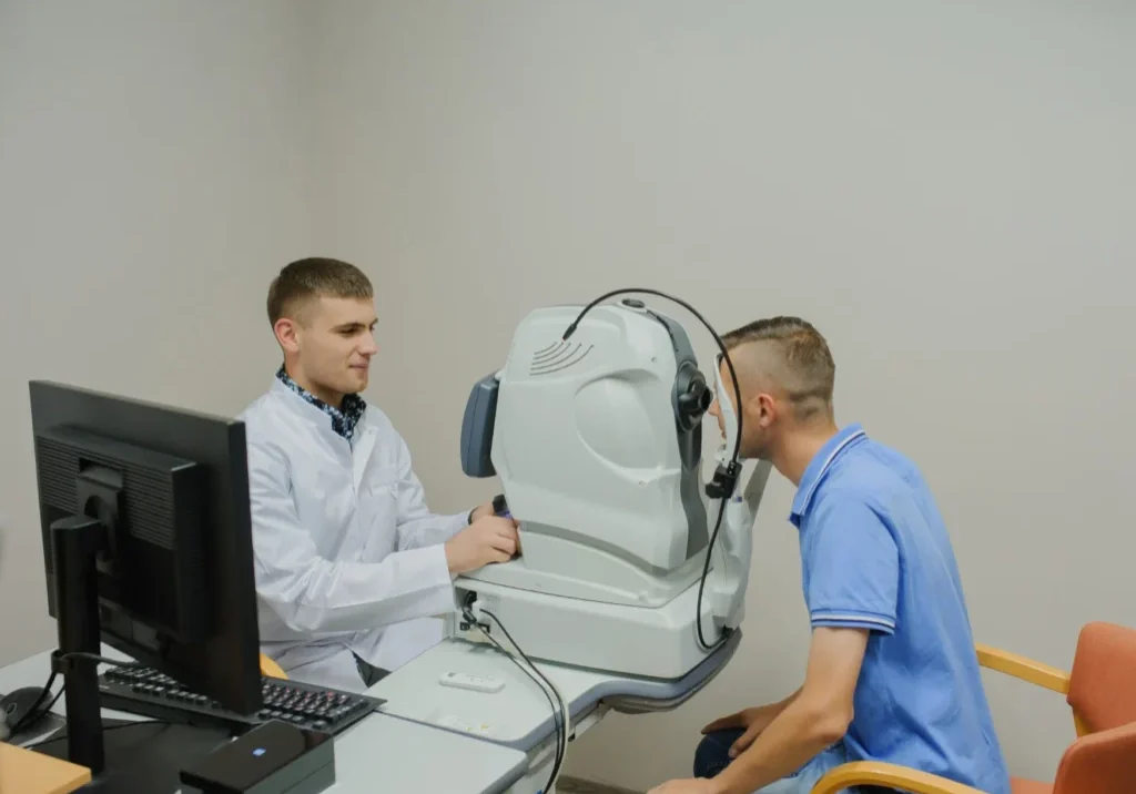
The patient sits at the machine, rests their chin on a support, and looks at a fixation target for a few seconds per eye. No dilation is required for most scans, no contact with the eye happens, and the whole process takes under five minutes for both eyes.
Behind that simplicity is a lot of engineering. The device sends a low-power light beam into the eye and measures how long it takes to reflect back from different tissue depths. That data builds a cross-sectional map, similar in concept to ultrasound but using light instead of sound waves, which is why resolution is so much finer: down to a few microns.
What the Scan Reveals
- Retinal nerve fiber layer thickness (key for glaucoma staging)
- Macular thickness and fluid buildup (key for wet AMD and diabetic macular edema)
- Optic nerve head structure
- Vitreoretinal interface issues, like epiretinal membranes or macular holes
Where OCT Fits in the Visit
Most practices run OCT as a screening step before the doctor exam, not as a replacement for it. The tech captures the scan, the software flags abnormal thickness patterns against a normative database, and the doctor reviews the images alongside the patient’s history. It’s a workflow multiplier: one machine, staffed by a technician, can process a full morning’s patient list without pulling the doctor away from the exam lane.
What Most Guides Miss: OCT Angiography Changes the Calculus
Most breakdowns of OCT stop at structural imaging. What they skip is OCT-A (OCT angiography), a newer capability on higher-end and recent-model machines that maps blood flow in the retina without dye injection. Traditional fluorescein angiography requires an IV and carries allergy risk. OCT-A gets similar vascular detail non-invasively, in the same scan session as the structural OCT.
For a practice weighing refurbished equipment, this is the detail that actually affects the purchase decision: not every OCT machine on the resale market has OCT-A, and not every practice needs it. A general optometry practice screening for glaucoma and routine macular disease often does fine without it. A practice managing a heavier retina caseload, or one taking referrals for diabetic retinopathy staging, will find OCT-A pays for itself in reduced referrals out.
If you’re comparing machines, our breakdown of what is an OCT machine and how the technology has evolved covers the generational differences in more detail.
Buying Considerations for Practices
A few things worth checking before signing off on any refurbished OCT purchase:
- Software update path. Older units sometimes stop receiving normative database updates, which limits diagnostic value over time.
- Scan speed. Faster scan rates mean fewer motion artifacts, especially useful with pediatric or unsteady patients.
- Integration with existing EHR and imaging systems. A machine that can’t export to your current workflow creates double documentation.
- Warranty and service support, particularly for refurbished units where parts availability varies by manufacturer.
- Axial resolution specs. Spectral-domain OCT units typically deliver resolution fine enough for most comprehensive practices. Swept-source models push finer and faster, but the price jump only makes sense if your caseload justifies it.
Training and Staffing Considerations
OCT is technician-operated in most practices, which means the learning curve matters as much as the hardware spec sheet. A tech who can capture a clean, well-centered scan on the first pass keeps patient flow moving; one who needs three or four attempts per eye adds real time to a busy schedule. Most manufacturers and refurbishment partners include operator training with a purchase, and it’s worth confirming that up front rather than assuming it’s included.
Image quality also depends on patient positioning and fixation, which is why a five-minute test scan during a practice visit or trial period tells you more than a spec sheet ever will. If a machine is hard to align on a wide range of patients, from cooperative adults to less steady seniors, that friction shows up every single day it’s in use.
We put together a full comparison in our guide to choosing the best OCT for your practice, which walks through specific models and where they land on price versus capability.
OCT is rarely purchased alone. Most practices pairing OCT with a corneal topographer get a fuller anterior-to-posterior diagnostic picture, and a biometer rounds out the pre-surgical workup for practices doing cataract referrals.
Final Thoughts on OCT Scan Machines
OCT scan machines have gone from a specialty retina tool to a standard part of the comprehensive eye exam in under two decades. The technology catches disease earlier, moves patients through the exam lane faster, and gives doctors a defensible record for every visit. Whether you’re outfitting a new practice or replacing an aging unit, the right OCT purchase comes down to matching capability to your actual patient mix, not buying the newest model on the shelf. If you want a second opinion on what fits your practice and your budget, reach out, and we’ll walk through it with you.